Powder Diffraction-Thin Film
Powder and Thin film studies with X-ray diffraction methods
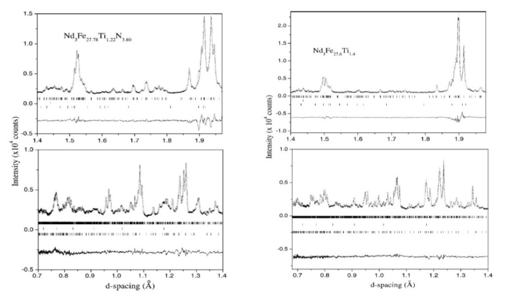
The X-ray powder diffraction technique (XRD) is used for the crystallographic study and characterization of polycrystalline materials of laboratory, industrial, geological, archaeological and pharmaceutical origin. Three diffractometers are available for polycrystalline samples and thin film characterizations. The materials are studied by using flat sample holders in D500-SIEMENS and Smart Lab Rigaku Diffractometers by applying the Bragg Brentano focusing geometry or using the Debye-Scherrer geometry with the Rigaku R-axis SPIDER single crystal diffractometer. The diffractograms in the last case is recorded on an Image Plate as 2D images (Debye-Scherrer rings). The SIEMENS D500 diffractometer was a key facility for the former Institute of Materials Science as its access was open to all users, an administration status which is still valid in the new Institute and the laboratory continues to offer its services to all the scientists and students of NCSR “Demokritos” and also to scientists from other research centers and Universities. During the 32 years of operation (it was installed at the end of 1989) approximately 100.000 samples have been measured. The XRD diagrams are analyzed using the Rietveld method for a complete crystallographic study of the sample under study or using database standards, for the identification of the crystalline phases contained in the studied sample. Almost all the studies in the area of powder diffraction have been done in collaboration with other groups of the Institute or with colleagues from Greek Universities or other Research Centers. The Rietveld method has been applied first for the study of hard-magnetic alloy materials in collaboration with the Pemanent Magnets, Magnetic Recording And Thermoelectrics Group. As an example we mention, the correct space group assignment to the structure of the hard magnetic phase Nd3 (Fe ,Ti)29 as a result of Rietveld analysis on X-ray diffraction data obtained with a conventional source [J. Μ. Μ. Μ 146 (1995) 335-345, doi:10.1016/0304-8853(94)01694-1 ]. The application of Rietveld-refinement method also on high-resolution Neutron Powder Diffraction data at room temperature for the parent and the nitrided compound [J. Phys.-Cond. Το Matter 15 (2003) 7953-7979, doi:10.1088/0953-8984/15/46/013 from our group gave an unambiguously determination of the space group and the calculated high-precision crystal structure parameters are used for the calculation of the corresponding Wigner-Size cell of each crystallographic site. The calculation of the polyhedral configuration of each site which is based on the nearest neighbours is a useful tool for comparative physical properties studies of structurally similar compounds.
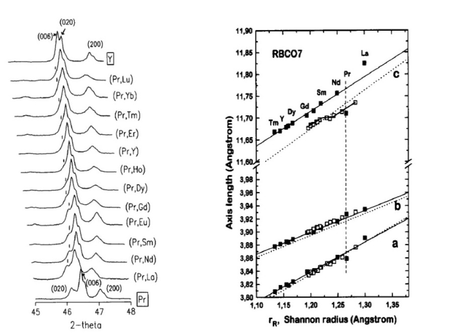
Another family of compounds which have been studied with powder diffraction methods and their structure has been analysed by using the Rietveld Method are oxides and especially those presenting superconducting properties in collaboration with Pemanent Magnets, Magnetic Recording And Thermoelectrics and Superconducting and Magnetic Oxides Groups and the IR Group of National Hellenic Research Foundation (collaboration with Dr. G. Chrisikos). As an example the two families of compounds RBa2Cu3O7 and [R0.5Pr0.5]Ba2Cu3O7 compounds with R=Y and lanthanides, are presented which have been studied at ambient temperature by means of XRD and infrared-reflectance spectroscopy Physica C 254 (1995) 44-62, doi:10.1016/0921-4534(95)00553-6].The interest for the study of these systems it is based on the fact that the compounds which contain Pr are not superconducting or the Tc transition temperature is reduced. In Pr free compounds, the change of the radius of lanthanides, leaves the puckering of the Cu(2)−O(2,3) sheet unaffected. In the Pr containing compounds, the Pr-O coordination polyhedron is typical of R+3 ions in eight-fold coordination and the observed changes argue against differentiating Pr in terms of valence or hybridization. Instead, a repositioning of Ba within the cuprate block is observed, leading to the flattening of the sheet and the anomalously high frequency of the Ba and Cu(2)−O(2, 3), B1u modes. In R-Pr compounds the c-axis and the interplanar distances related to the position of Ba and O(2, 3) are found to exhibit a sigmoidal dependence on rR. Based on the present study it has been argued that this sigmoidal dependence is related to the two-mode behavior exhibited by several phonon modes of these compounds.
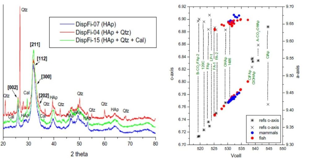
Conserning the study of samples of archaeological origin, a systematic study published in [Palaeogeography, Palaeoclimatology, Palaeoecology 534, άρθρο #109334 (2019), doi:10.1016/j.palaeo.2019.109334] in collaboration with the Department of Historical Geology & Palaeontology, Faculty of Geology & Geoenvironment, of the University of Athens aimed at identifying the nature and origin of “black” bones from Dispilio, based on the hypothesis of a fire-destruction event, indicated by other lines of evidence (micromorphology, palaeobotanical study). The use of fire within a prehistoric settlement is one of the major issues of archaeological research as it proves the presence of man. Concerning the mineralogical content, the XRD analysis give that the main phase in all studied samples is bioapatite but the mineral phases of calcite and quartz are also present. Based on the broad peaks exhibited by our samples in the XRD patterns, as well as the Crystallinity Index (C.I.) values calculated, the bioapatite of our samples seems to be poorly crystalline as the C.I. to take values in the range 0.07-0.16 for the studied samples. The crystal size along the c-axis was estimated through the Rietveld analysis to be around 110 Å, supporting the hypothesis of poor crystallinity and the samples did not exhibit any grouping based on crystallinity. The latter means that the crystallinity of the Dispilio samples has not been significantly affected due to recrystallization and the thermal alteration of bone is known to affect the crystallinity of bioapatite. Burned samples give values for C.I. in the range of 0.2-1.2. The distribution of the studied samples agrees with the calculated crystallinity indices, as the most crystalline of our samples are closest to the fluorinated apatites. It is known that the lower the Vcell and a-axis values are, the larger the crystal size of apatite’s are. Concluding the various analytical techniques employed on mammal and fish bones did not offer any proof that any of the studied material is burnt, based on specific features described in relevant bibliography. Rather, all the observed alterations concerning cracking, histology, color, crystallinity etc. in the samples, could be attributed to diagenesis.
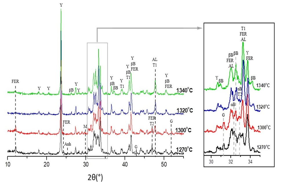
The study of Industrial samples concerns the research on alternative cement clinkers which have been proposed as building materials with improved carbon footprint compared to conventional Portland Cement (PC) whose production is a highly energy-intensive industrial process. In a study [CEMENT AND COCONRETE RESEARCH, 147 (2021)106529), doi:10.1016/j.cemconres.2021.106529] performed in collaboration with the Group Ceramics and Composite materials of INN, the effect of clinkering temperature and duration on the production of low energy Belite Calcium-Sulpho-Aluminate (BCSA) clinkers, targeting a composition belite and ye’elimite at 40 wt. % each was examined . The mineralogy and microstructure of BCSA clinker were studied by XRD/QXRD (Figure-4) and SEM/EDS analysis. The target concentration of belite was achieved at temperatures between 1270 and 1300 oC, while that of ye’elimite was only marginally reached at 1300 and even at 1320 oC. In all clinkering conditions belite was detected in both αH’- and β- polymorphs. The extensive use of bauxite to produce BCSA clinker lead to a Ti - rich raw mix and the formation of perovskite in all clinkers. At low clinkering conditions the presence of Ti and S-ions in belite crystals contributed to the stabilization of belite αH’- polymorph, while at elevated temperatures their partial release from belite crystal favors the stabilization of β - polymorph.
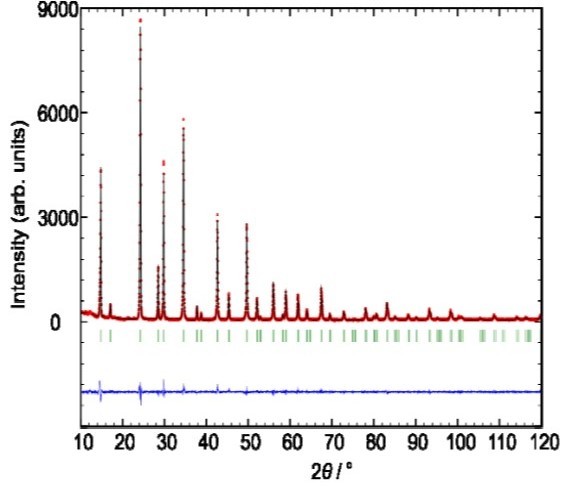
In collaboration with the group Nanotechnology Processes for Solar Energy Conversion and Enviromental Protection of INN the structure, the vibrational and the optical properties of the defect perovskites Cs2SnX6 (X = Cl, Br, I) have been studied [The Journal of Physical Chemistry C 120 (2016) 11777-11785, doi:10.1021/acs.jpcc.6b02175]. A rietveld analysis plot for the Cs2SnCl6 compound is given bellow.
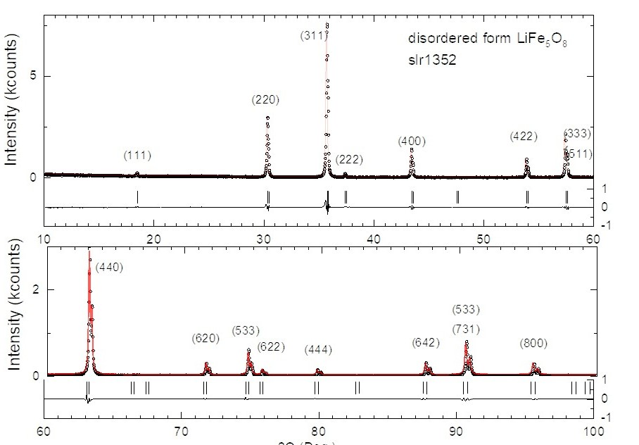
Recently, the Lithium Ferrite oxides are studied in collaboration with the Superconducting and Magnetic Oxides Groups Group, as this materials are used for cathode materials in Li-ion batteries, microwave magnetic applicatiosns etc. In following Figure, a representative rietveld refinement diagram of the disordered LiFe5O8 phase with spinel structure which crystallizes in the space group F d-3 m, is given.
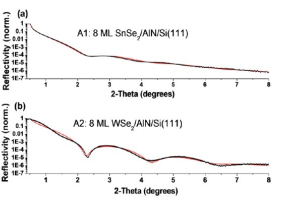
The Smart Lab diffractometer is equipped with the CBO unit which produces parallel beam and this setup is used for X-ray reflectivity measurements and Grazing incidence X-ray diffraction. In the following Figure the XRR measurement and analysis of thin films from SnSe2 and WSe2 which gave a thickness of 3.7 and 4.2 nm respectively, are presented [ACS Appl. Mater Interfaces 8 (2016) 23222-23229 doi:10.1021/acsami.6b02933]. The analysis was performed for the needs of the Graphene and two Dimentional Materials for Nanoelectronics group.
During the last decade, the laboratory has carried out a large number of measurements on samples from industries and analytical chemical laboratories. The main colaborations based on contracts are with the quality control and R&D departments of PHARMATEN Pharmaceutical Industry A.B.E.E. and PHARMATHEN INTERNATIONAL S.A. as well as with BIC Violex ABEE. The income from the services provided are used to maintain facilities, to pay staff, and purchase equipment and programs.