Physics of Oxides
A systematic study on ferrimagnetic LiFe5-xMnₓO₈ spinel is conducted using X-ray diffraction, Mössbauer spectroscopy, and magnetic measurements. We investigated how different synthesis conditions influence the crystal chemistry and magnetic properties of this compound, a crucial step towards its potential applications in microwave, spintronic, and electromagnetic devices, as well as in lithium-ion battery cathodes. Our results show a method for reversibly producing composite (two-phase) materials with comparable crystal and magnetic properties. We uncovered a fine phase separation in the post-annealed samples. This phase separation arises from manganese substitution for the iron, which prevents the lithium ordering seen in the pure post-annealed LiFe₅O₈ compound. The two phases observed have the same spinel structure and nearly identical magnetic properties, but slightly different unit-cell constants. From hyperfine parameters of iron extracted from Mössbauer spectra, we found that iron in all samples is in a trivalent (III) high-spin (S=5/2) state. Based on the observed spectral area ratio of two sextets in MS and the saturation magnetization values at liquid-helium temperatures, a model for the distribution of cations between tetrahedral and octahedral sites is proposed. Understanding the crystal chemistry of these materials could be beneficial for their applications in microwave, spintronic, and electromagnetic systems, as well as in lithium-ion battery cathodes.
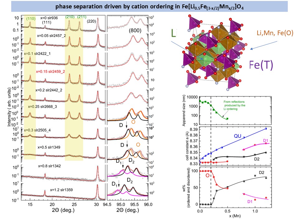
Evolution of the Rietveld plots of the post-annealed LiFe5-xMnₓO₈ samples. For x<0.25, the ordered LiFe5O8 compound is observed with decreasing apparent size (see right panel). For x>0.25, two phases are observed with the disorder LiFe5O8 spinel structure.
For more details, see: https://doi.org/10.1557/s43578-026-01794-w
Exploring Hidden Structures in LiFeO₂: Insights from Mössbauer Spectroscopy and X-Ray Analysis
Binary oxides like LiMO₂ (where M = Ti, Mn, Fe, Ni, or Co) are attracting significant attention for their versatile crystal structures and promising applications—especially as cathode materials in lithium-ion batteries. Among them, LiFeO₂ stands out as a closely related compound to those already used in battery technologies.
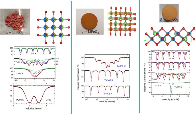
To deepen our understanding of how this material behaves, we investigated the crystal and magnetic properties of polymorphic LiFeO₂ using Mössbauer spectroscopy, X-ray diffraction (XRD), and magnetic measurements. Our findings reveal a complex and fascinating picture: (a) Short-range structural ordering in the α-LiFeO₂ phase appears to be connected to embedded nanoregions of β′-LiFeO₂. (b) Low-temperature Mössbauer spectra reveal a disordered magnetic state in α-LiFeO₂ below 90 K. (c) Rietveld refinement of the γ-LiFeO₂ phase indicates a defect-rich microstructure, providing insights into nanoscale imperfections. While isolating pure β′-LiFeO₂ remains a challenge, we found that annealing α-LiFeO₂ at 400°C creates a nanocomposite featuring both β′-LiFeO₂ and γ-LiFeO₂ nanoregions. These findings bring us a step closer to understanding how nanoscale structures influence the performance and stability of battery materials, a crucial insight for the next generation of energy storage systems.
For more details, see: https://doi.org/10.1557/s43578-024-01338-0
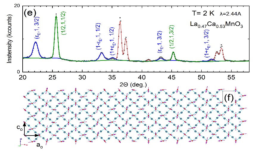
Mixed orbital states and modulated crystal and magnetic structures in La1-xCaxMnO3 deduced from synchrotron X-ray and neutron diffraction data.
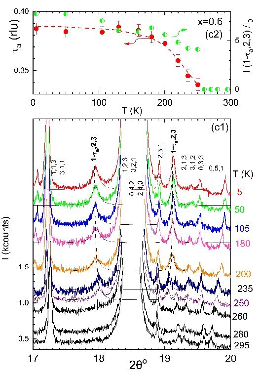
Several important phenomena, including ferromagnetic magnetoresistance effects, charge, and orbital-ordered states, have been observed in the model-doped La1-xCaxMnO3 manganese perovskite. We have studied the crystal structure in a series of compounds with 0.5< x≤0.6 using ultrahigh-resolution synchrotron X-ray (see figs. C1&c2) and neutron (see figs. e&f) diffraction data in high-quality polycrystalline samples. The experimental results reveal that all compounds undergo a structural transition at T<TCO(x)~200-220 K with the concomitant emergence of superlattice Bragg peaks, which can be indexed assuming a superstructure with a modulation propagation vector, τ. At the base temperature of 5 K, the modulation vector of the superstructure τ=[τa,0,0] is parallel to the a-axis, with τa varying linearly with x, as τa~1-x. A sinusoidal modulated structure has been proposed to interpret the neutron diffraction data. This result may be linked to a mixed orbital state of the manganese ions. Our findings may be useful for understanding phenomena related to spin, charge, and orbital ordering in greater depth, as well as symmetry breaking and emergent order in quantum states [1,2].
Manganese Perovskites
Manganese perovskites with the general chemical formula La1-xCaxMnO3 have been thoroughly studied by us in the past using magnetic, NMR, EPR, Mossbauer, X-ray, and neutron data. In this system, several important, poorly understood issues stay, related to the physics of strongly correlated electron systems. The following shows a representative specific-heat measurement to elucidate the nature of the phase transitions observed in the diffraction and magnetization studies. Our measurements have focused on the ferromagnetic-insulating regime of the magneto-structural phase diagram. Figure 1 shows the magneto-structural phase diagram, which we have estimated using neutron and synchrotron X-ray diffraction data. The left panel of Figure 1 shows a representative specific-heat measurement for the sample with x=0.15. The data clearly show a λ-type second-order transition, which coincides with the paramagnetic-canted antiferromagnetic transition detected in magnetization and neutron diffraction data. At room temperature, a symmetric peak is seen, arising from the Jahn-Teller structural transition. The symmetric peak shape implies a first-order structural phase transition. The anomaly observed around liquid N2 temperatures in the magnetic measurements is absent in the specific heat measurements.
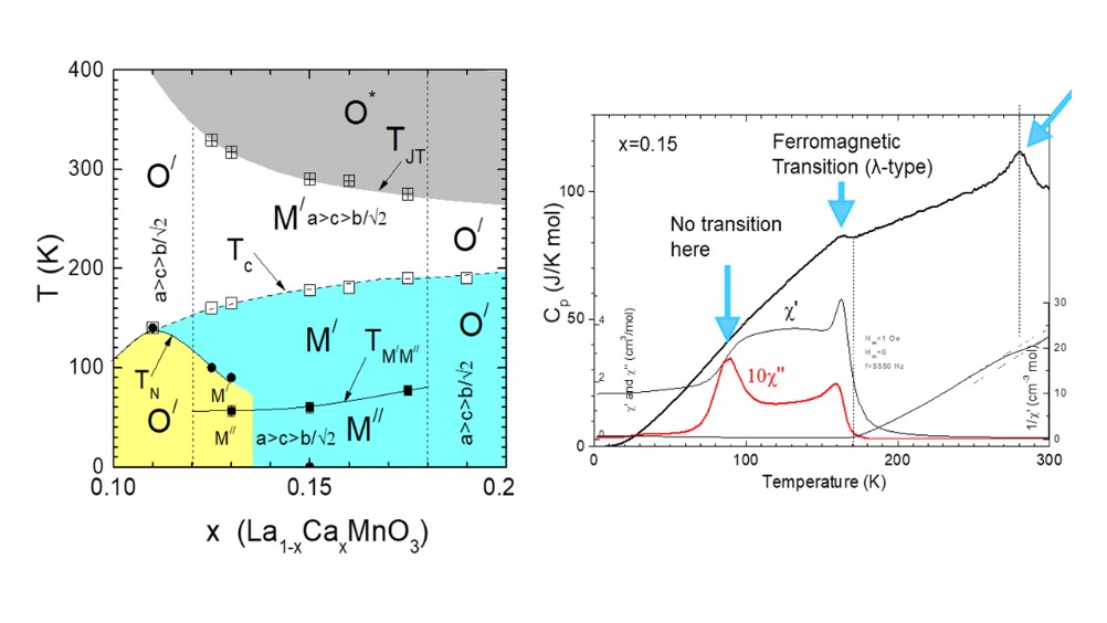
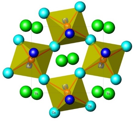
Fig. 1 (left upper panel) Part of the magneto-structural phase diagram of the La1-xCaxMnO3 compound in the canted antiferromagnetic -ferromagnetic insulated regime. (right upper panel) Specific heat measurements of the La1-xCaxMnO3 compound, with x=0.15. (lower panel) Unit cell of La1-xCaxMnO3 compound
Ferrites for microwave and 5G communication systems
Ferrites like Y3Fe5O12, LiFe5O8, is one of the few ferrimagnetic insulators with low microwave losses and high saturation magnetization and Curie temperatures. These properties may be useful in the construction of ultra-wideband photonic devices for 5G communication systems.
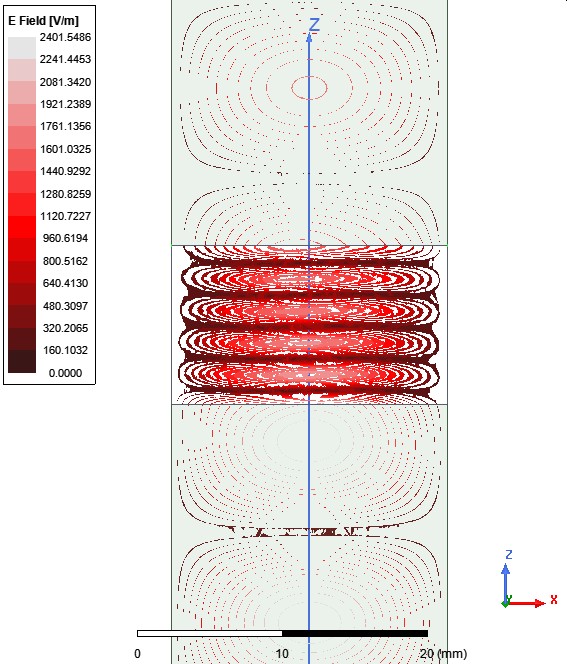
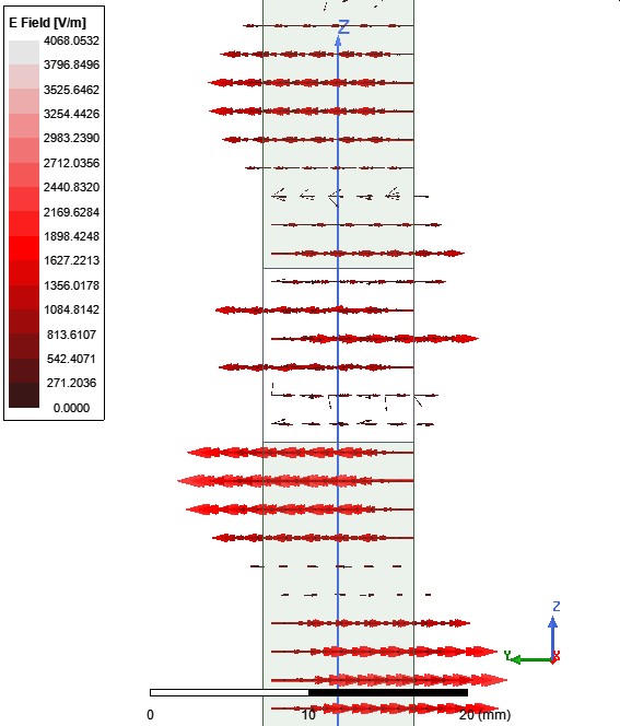
Fig. 2 Contour and vector plot of the electric field on the surface y = b / 2 of the wave guide, loaded with Y3Fe5O12 ferrite, under a magnetic field Hy = 5.55 kOe, for time phase 0o.The frequency of the ac-electric field was f=14.3 GHz and corresponds to the frequency where the third peak of the transmission coefficient S21 occurs. The ac-electric field was estimated using ANSYS HFSS (2019 R3).
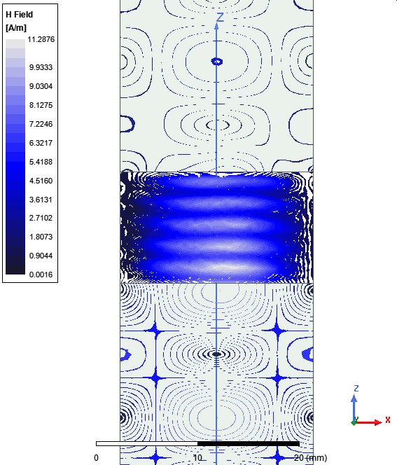
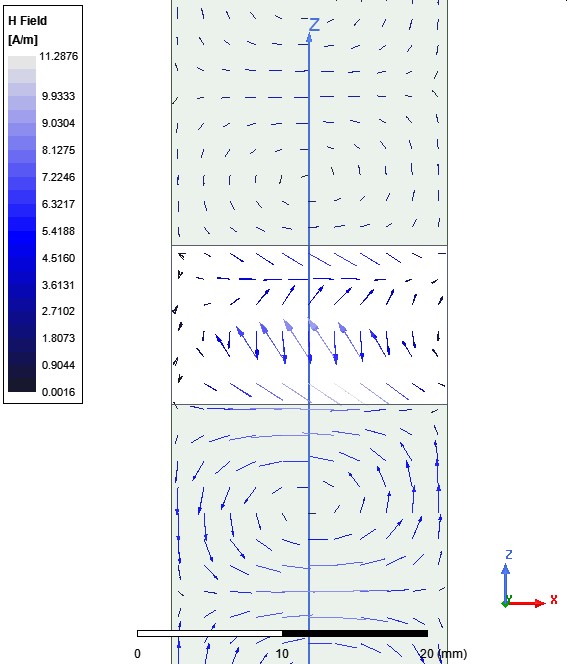
Fig. 3 Contour and vector plot of the magnetite field on the surface y = b / 2 of the wave guide loaded with Y3Fe5O12 ferrite, under a magnetic field Hy = 5.55 kOe for time phase 0o. The frequency of the ac-magnetic field is f=14.3 GHz and corresponds to the frequency where the third peak of the transmission coefficient S21 occurs. The ac-magnetic field was estimated using ANSYS HFSS (2019 R3).
To design these devices, it is necessary to know with high accuracy the material parameters of the ferrites, such the electric permittivity and the parameters of the magnetic permeability tensor. One of the used methods, is the measurement of the scattering parameters of a wave guide filled with a magnetized ferrite. Since it is impossible to calculate theoretically the scattering parameters (S11, S12) and with a fitting procedure to calculate the material parameters, is plausible to resort to a method where the theoretical scattering parameters will be calculated by the numerical solution of the Maxwell equations. To this end we used the ANSYS electromagnetic suite of programs (particularly HFSS, version 2019R3), to calculate the scattering parameters (see fig 2 and 3). By employing a trial-and-error methodology we succeeded to reproduce the experimental scattering parameters, using the same electromagnetic materials parameters for all values of the magnetic field. After few trial simulations we found out that we can have an accepted coincidence between theoretical and experimental scattering parameter, when Oe (see fig4)
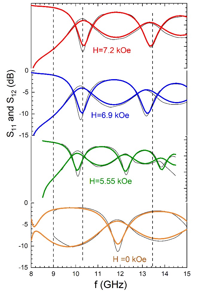
Fig. 4 Experimental (thin black lines) and theoretical calculated from the simulation (thick colored lines) scattering parameters S11 and S21 of Y3Fe5O12 ferrite.
Magnetoelectric oxides
The coupling between magnetic and (ferro)electric order is called the magnetoelectric effect, which is the induction of magnetization by an electric field or the induction of electric polarization by a magnetic field. The magnetoelectric effect has been studied theoretically in the past by Curie and Dzyaloshinskii, and experimentally by Astrov. Magnetoelectric materials can be used in novel devices, such as computer memories, where magnetic (ferroelectric) domains can be controlled by an electric (magnetic) field, sensors, and spintronic devices. The magnetoelectric behavior in existing materials occurs at low temperature, and the elements of the magnetoelectric tensor are very small, limiting their practical use.
YBaCuFeO5 compound is an antiferromagnetic insulator with Neel temperature around TN=400 K. Below TN2 (depending on the preparation conditions, see Fig. 3), the magnetic structure changes from a colinear simple antiferromagnetic to an incommensurate magnetic structure (a circular inclined spiral with propagation vector k=(1/2,1/2,1/2 ± q). More importantly, an electrical polarization appears below TN2, which scales with the modulus of the magnetic modulation vector q down to the lowest temperature. Our research on this topic focuses on studying the physical origin of the magnetoelectric effect using Mossbauer spectroscopy. Fig. 5 shows representative Mossbauer spectra from two YBaCuFeO5 samples with different incommensurate magnetic transitions.
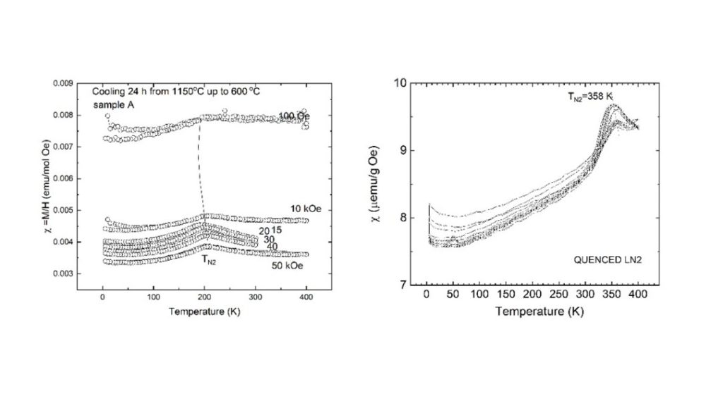
Fig. 5 Temperature variation of the magnetic dc susceptibility of two YBaCuFeO5 samples produced by slowly cooling from 1150 °C and quenching for 1150 °C to LN.
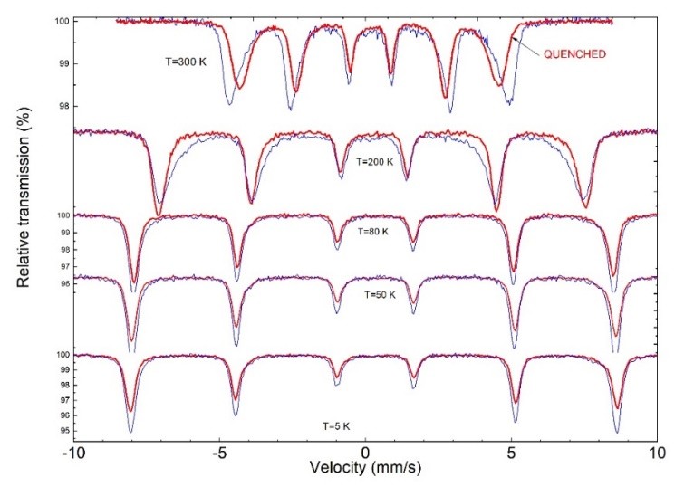
Fig. 6 Mossbauer spectra of the YBaFeCuO5 compound produced with slow cooling (blue spectra) and quenched from 1150 °C to LN (red spectra).
The physical origin of the magnetoelectric properties of the Y3Fe5O12 ferrimagnetic compound
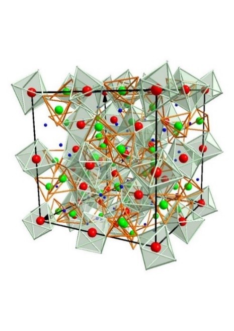
Fig. 7 Unit cell of Y3Fe5O12. YIG crystallizes in the cubic system (space group Ia-3d)., The unit cell contains eight formula units. Oxygen anions occupy the general position 96h (x, y, z), Y the dodecahedral 24c (1/8,0,1/4) sites, and Fe occupy two non-equivalent sites [16a (0,0,0) and 24d (3/8,0,1/4)] with octahedral and tetrahedral coordination, respectively.
Y3Fe5O12 (YIG) is a ferrimagnetic prototype compound exhibiting interesting physical properties (see Fig. 7). It has been used in several practical applications, such as magnetooptical sensors and microwave ferrites. In recent years, extensive studies have focused on the spin-wave decay length of thin films of YIG. It was determined to be several centimeters, thus allowing the spin angular moment to propagate over relatively long distances.
This makes YIG an ideal conductor for spin currents transmitted via spin-wave excitations. In combination with the spin Hall or inverse spin Hall effect, thin-film structures based on YIG could provide a new method for transferring spin information via spin-wave excitations. Another important aspect of YIG concerns its magnetoelectric and magneto-capacitive properties, which are targeted for applications in novel microwave antennas.
The magnetoelectric (ME) coupling concerns either a variation in the dielectric response upon application of a variable external magnetic field, or a variation in the magnetic response due to a variation in the electric field. The ME properties of YIG have long been debated, and its microscopic origin remains a long-standing controversy in the scientific community. The controversy relies on the physical requirement that permanent electrical polarization can be observed in materials with a non-centrosymmetric crystal structure. So far, all reported crystallographic data on purely stoichiometric YIG do not provide experimental evidence for a non-centrosymmetric center.
X-ray linear dichroism data have been reported for epitaxially grown thin films of YIG, which are consistent with small structural distortions that make rhombohedral subgroups more plausible, suggesting either space group R-3 in the ferrimagnetic phase or R3m in the magnetoelectric phase. To elucidate the complex behavior of YIG it is required to understand the puzzling effect of dispersion in electric permittivity and magnetic susceptibility. Our current effort focuses on the dielectric and magnetic relaxation effects of YIG samples with different microstructures, employing Mossbauer spectroscopy, X-ray diffraction, scanning electron microscopy, specific heat, magnetic, and dielectric measurements. The results show that dielectric and magnetic relaxation processes occur only in samples prepared by a solid-state process in our laboratory.
Fig.8 displays the surface morphology observed by SEM on the free surface of pellets from three YIG samples. The first figure corresponds to a commercial sample and displays low dielectric and magnetic losses. The center and the right picture show the surface morphologies of two YIG samples prepared in our laboratory at 1400oC and 1500oC respectively. The sample prepared at 1400 °C displays significant dielectric and magnetic losses as well as magnetoelectric behavior. Interestingly, in the sample prepared at 1500 °C, the electric and magnetic losses are absent. The surface morphology of the commercial sample reveals polygonal grains of various sizes, with negligible porosity. The other two samples display significant porosity. The sample prepared in 1500oC displaying a second phase at the grain boundaries.
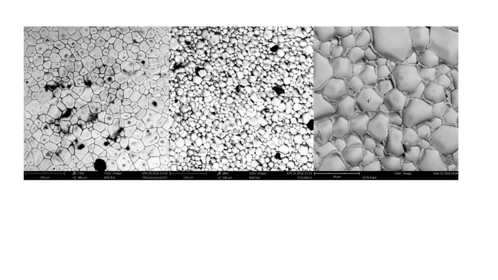
Fig. 8 Scanning electron photographs of three YIG samples prepared under different conditions. (left photo) Commercial sample. (center) a YIG sample sintered at 1400 °C and (right) a YIG sample sintered at 1500 °C.
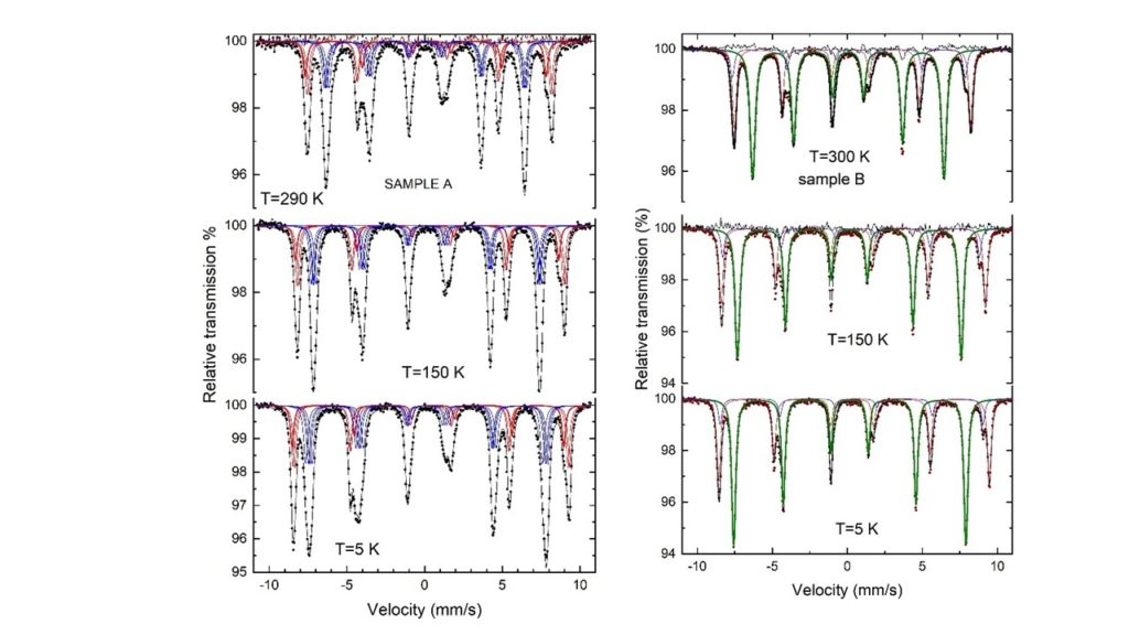
Fig. 9 (left) Mossbauer spectra of the commercial YIG sample. (right) Mossbauer spectra of our sample prepared at 1400oC.
Fig. 9 shows Mossbauer spectra (MS) collected at 295 K, 150 K, and 5 K for the commercial and the YIG samples prepared in our laboratory. These spectra can be analyzed with three partially resolved sextets. The sextet with a lower hyperfine magnetic field is assigned to the tetrahedrally coordinated iron Fe+3, whereas the other two sextets exhibit similar hyperfine magnetic fields and are assigned to octahedrally coordinated Fe+3. Octahedral Fe exhibits two different sextets because each octahedral site has its principal axis of the electric field gradient (EFG) along one of the [111] crystallographic directions, which are the easy directions of magnetization, and the MS are split into two sextets corresponding to two different mutual orientations between the internal field and the principle of the EFG tensor. These two mutual orientations between the EFG z-axis and the direction of the hyperfine magnetic field [111] correspond to either an angle Θ=70o or 0 °. Thus, the two sextets from octahedral sites can be assigned to nearly similar values of hyperfine magnetic field H. For the tetrahedral site, the angle between the principal axis of the EFG sensor and the hyperfine magnetic field is 54 °. The main difference between the commercial sample and our sample is in the pronounced broadening of the tetrahedral component. The additional line broadening observed in the MS of the commercial sample was modeled with four sextets with equal magnetic hyperfine field, isomer shift, and principal value of the EFG sensor, but different angles between the z-axis of the EFG sensor and the hyperfine magnetic field (parallel to the magnetization). This complication strongly suggests that this sample is either non-stoichiometric or that the crystal symmetry becomes rhombohedral. The sample prepared in our laboratory can be fitted with three relatively narrow sextets, implying that it is stoichiometric, or the rhombohedral distortion is very small.