3D Scaffolds for tissue engineering
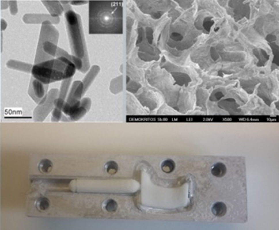
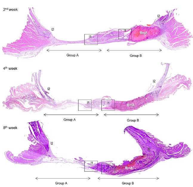
Our work is focused on the development of hydroxyapatite nanoparticles using dendritic polymers as biomimetic templates. Following their successful preparation,1,2 hydroxyapatite nanoparticles were employed for the development of biopolymer/nanohydroxyapatite porous personalized 3D bio-implants of complex geometry.3 A number of studies were performed providing evidence that these scaffolds exhibit increased biocompatibility and bioactivity and are able to promote guided bone regeneration in rat calvarial critical-sized defects.4-6
The work was partially supported by a national project.
Relevant Publications
- D. Tsiourvas, A. Tsetsekou, M.-I. Kammenou, N. Boukos, Controlling the formation of hydroxyapatite nanorods with dendrimers, J. Am. Ceram. Soc., 94, 2023–2029 (2011).
- D. Tsiourvas, A. Tsetsekou, M.-I. Kammenou, N. Boukos, Biomimetic synthesis of ribbon-like hydroxyapatite employing poly(L-arginine), Materials Science and Engineering C, 58, 1225–1231 (2016).
- D. Tsiourvas, A. Sapalidis, T. Papadopoulos, Hydroxyapatite/chitosan-based porous three-dimensional scaffolds with complex geometries, Materials Today Communications 7, 59–66 (2016).
- E. Chatzipetros, S. Damaskos, K. I. Tosios, P. Christopoulos, C. Donta, E.-M. Kalogirou, Z. Yfanti, D. Tsiourvas, A. Papavasiliou, K. Tsiklakis, The effect of nano-hydroxyapatite/chitosan scaffolds on rat calvarial defects for bone regeneration, International Journal of Implant Dentistry, 7, 40 (2021); doi:10.1186/s40729-021-00327-w.
- E. Chatzipetros, Z. Yfanti, P. Christopoulos, C. Donta, S. Damaskos, E. Tsiambas, D. Tsiourvas, E.-M. Kalogirou, K. I. Tosios, K. Tsiklakis, Imaging of nano-hydroxyapatite/chitosan scaffolds using a cone beam computed tomography device on rat calvarial defects with histological verification, Clinical Oral Investigations, 24, 437–446 (2020); doi:10.1007/s00784-019-02939-4.
- E. Chatzipetros, P. Christopoulos, C. Donta, K. I. Tosios, E. Tsiambas, D. Tsiourvas, E.-M. Kalogirou, K. Tsiklakis, Application of nano-hydroxyapatite/chitosan scaffolds on rat calvarial critical-sized defects: A pilot study, Med. Oral Patol. Oral Cir. Bucal. 23, e625–632 (2018); doi:10.4317/medoral.22455.